Skip to content
Home
About Us
Our Services
ACUPUNCTURE AND ELECTROTHERAPY
Cardiac Surgery
Endoscopy
LASER THERAPY AND PHOTO DYNAMIC THERAPY
NUTRIGENOMICS AND NUTRACEUTICALS
Orthopedic Surgery
Oncology
Animal Communication & Counselling
GERONTOLOGY
Advanced Diagnostic Imaging
OWNER ACCOMPANIED INDIVIDUAL RECOVERY SUITES
ICU and Negative Pressure Wards
Exotic Animals
AQUACULTURE CONSULTATION
Ozone Therapy
Microsurgery
Reconstructive Surgery
Bioresonance Allergy Testing
Cryosurgery
Dermatology
CANCER – IMMUNE CELL THERAPY & CELL BANKING
REGENERATIVE MEDICINE (IMMUNE CELL AND STEM CELL THERAPIES)
HYPERBARIC OXYGEN IN WOUND HEALING
Neuro Surgery
Ophthalmology
Full Blood Diagnostics
Pet Cloning
Physiotherapy
Our Team
Latest Updates
Contact Us
Career
SHOP
Main Menu
Home
About Us
Our Services
Menu Toggle
Advanced Diagnostic Imaging
Neuro Surgery
Endoscopy
Microsurgery
Orthopedic Surgery
Ophthalmology
Cardiac Surgery
Reconstructive Surgery
Cryosurgery
ICU and Negative Pressure Wards
OWNER ACCOMPANIED INDIVIDUAL RECOVERY SUITES
GERONTOLOGY
Oncology
Exotic Animals
Dermatology
Full Blood Diagnostics
REGENERATIVE MEDICINE (IMMUNE CELL AND STEM CELL THERAPIES)
CANCER – IMMUNE CELL THERAPY & CELL BANKING
NUTRIGENOMICS AND NUTRACEUTICALS
ACUPUNCTURE AND ELECTROTHERAPY
Bioresonance Allergy Testing
Pet Cloning
LASER THERAPY AND PHOTO DYNAMIC THERAPY
Physiotherapy
Animal Communication & Counselling
HYPERBARIC OXYGEN IN WOUND HEALING
AQUACULTURE CONSULTATION
Ozone Therapy
Our Team
Latest Updates
Contact Us
Menu Toggle
Career
SHOP
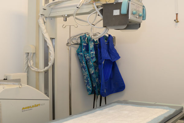
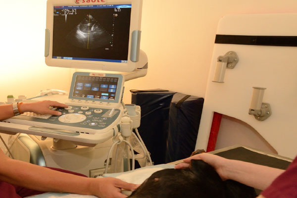
Veterinary Diagnostic Imaging
Know More About Veterinary Diagnostic Imaging
ADVANCED DIAGNOSTIC IMAGING (DIGITAL RADIOGRAPHY, ULTRASONOGRAPHY)
Scroll to Top